Embark on a journey of anatomical exploration as we delve into pal cadaver appendicular skeleton pectoral girdle lab practical question 6. This comprehensive guide unravels the intricacies of the appendicular skeleton, providing a hands-on approach to understanding its structure and clinical significance.
Through a meticulous examination of cadaveric specimens, you will gain an intimate knowledge of the bones, joints, and anatomical landmarks that define the appendicular skeleton. Our exploration extends beyond mere identification, delving into the functional roles and clinical applications of this essential skeletal system.
Appendicular Skeleton

The appendicular skeleton comprises the bones of the limbs and the girdles that connect them to the axial skeleton. It plays a crucial role in supporting the body, providing mobility, and protecting vital organs.
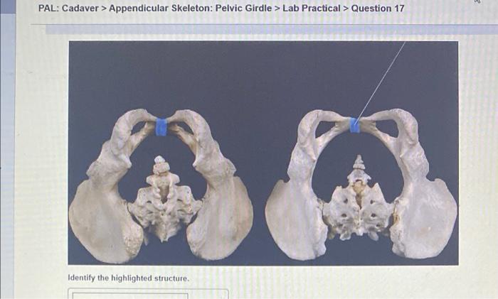
The appendicular skeleton can be divided into two main regions: the pectoral girdle and the pelvic girdle. The pectoral girdle supports the upper limbs, while the pelvic girdle supports the lower limbs.
Pectoral Girdle, Pal cadaver appendicular skeleton pectoral girdle lab practical question 6
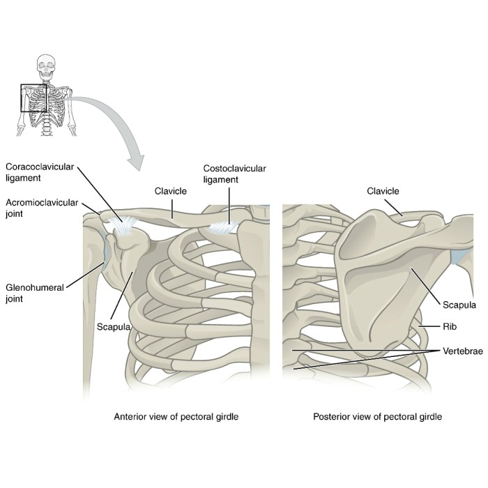
The pectoral girdle consists of the clavicle and the scapula. The clavicle is a slender bone that connects the sternum to the scapula. The scapula is a triangular bone that forms the shoulder blade.
The pectoral girdle provides a stable base for the upper limbs and allows for a wide range of movements, including flexion, extension, abduction, adduction, and rotation.
Appendicular Skeleton Bones
The appendicular skeleton consists of the following bones:
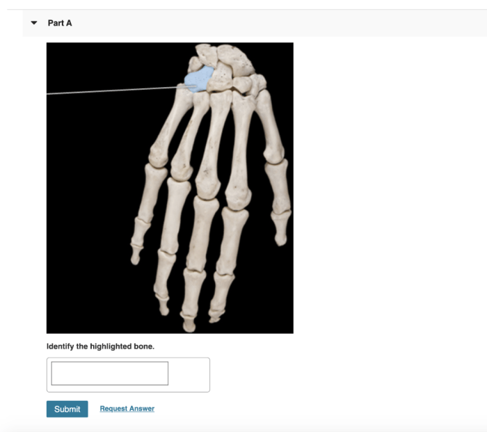
- Upper limbs: humerus, radius, ulna, carpals, metacarpals, phalanges
- Lower limbs: femur, tibia, fibula, tarsals, metatarsals, phalanges
Appendicular Skeleton Joints
The appendicular skeleton contains various types of joints, including:
- Ball-and-socket joints: allow for a wide range of movements, such as flexion, extension, abduction, adduction, and rotation (e.g., shoulder joint)
- Hinge joints: allow for flexion and extension (e.g., elbow joint)
- Pivot joints: allow for rotation (e.g., radioulnar joint)
- Condyloid joints: allow for flexion, extension, abduction, and adduction (e.g., wrist joint)
Clinical Significance of Appendicular Skeleton
Understanding the appendicular skeleton is essential for diagnosing and treating injuries or disorders of the limbs and joints. For example, knowledge of the anatomy of the shoulder joint is crucial for diagnosing and treating shoulder dislocations or rotator cuff tears.
Lab Practical Question 6: Palpation of Appendicular Skeleton
Palpation is a technique used to identify and examine the bones of the appendicular skeleton.
To palpate the bones of the upper limb:
- Clavicle: Palpate along the anterior surface of the shoulder from the sternum to the acromion process.
- Scapula: Palpate the spine of the scapula, acromion process, and inferior angle.
- Humerus: Palpate the shaft of the humerus, lateral and medial epicondyles, and trochlea.
- Radius and ulna: Palpate the styloid processes of the radius and ulna.
To palpate the bones of the lower limb:
- Femur: Palpate the greater trochanter, femoral shaft, lateral and medial condyles.
- Tibia and fibula: Palpate the tibial tuberosity, medial malleolus, and lateral malleolus.
Query Resolution: Pal Cadaver Appendicular Skeleton Pectoral Girdle Lab Practical Question 6
What is the significance of the appendicular skeleton?
The appendicular skeleton provides support and mobility to the upper and lower limbs, enabling a wide range of movements essential for daily activities.
How many bones make up the appendicular skeleton?
The appendicular skeleton consists of 126 bones, including the bones of the upper and lower limbs, the pectoral girdle, and the pelvic girdle.
What are the different types of joints found in the appendicular skeleton?
The appendicular skeleton contains various types of joints, including ball-and-socket joints, hinge joints, pivot joints, and gliding joints, each allowing for specific types of movement.

